Why 3D CT Imaging Matters
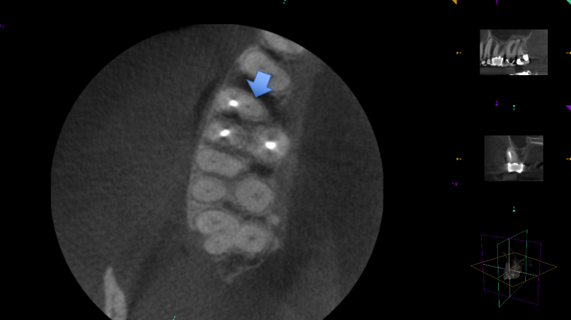
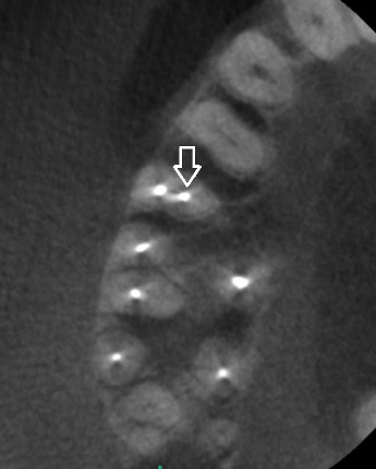
In our office we use advanced 3D imaging to find and clean out all of the infected tissue inside the tooth. The goal for a successful root canal is to remove all of the soft tissue that is like skin on the inside of the tooth.
The tricky part is that no two teeth are exactly alike. The same exact tooth in one person can have one root and in another person three roots.
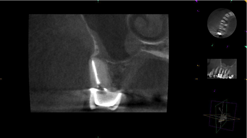
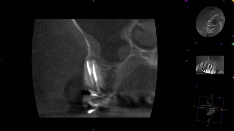
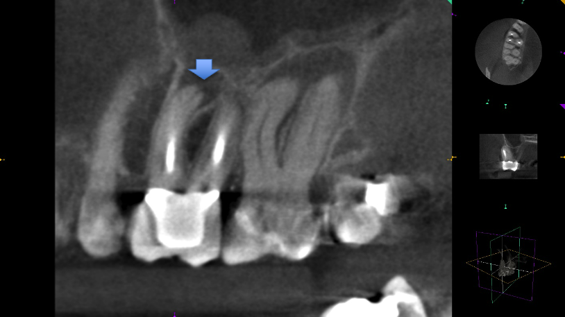
Old-school, flat dental X-rays simply can’t show tiny hidden details inside a tooth, but the same CT technology that doctors use in hospitals is now available in dentistry.
We’ve been using 3D CT machines since 2012—our office was the first in the area. This technology gives Doctor Bernstein a crystal-clear map of your tooth, making it much easier to find every bit of infected tissue to ensure your root canal is a success.
Why Root Canals Fail
If a tiny spot of infected tissue gets missed in a root canal procedure, bacteria will keep feeding on it. Eventually those bacteria will travel out through an opening at the bottom of the tooth’s root and into your jawbone. From there, they can jump into your bloodstream and travel to other parts of your body.
Because of this, we strongly encourage all of our patients to come back one year after their treatment for a quick follow-up 3D CT scan of their jaw. This lets us make sure the bone is healing perfectly and the infection is completely gone.
Here is the most important thing to know: teeth can be infected without causing any pain. You could chew your food perfectly fine and feel zero discomfort, while still having a serious infection hidden in your jaw.
If you skip your follow-up checkup, those hidden bacteria can leak into your blood, which can increase your risk for major health issues like heart disease and stroke. Since normal dental X-rays usually miss these quiet infections, a 3D CT scan is the best way to be absolutely sure you are healthy.
Getting Ready for Heart Surgery, Chemotherapy, Sinus Surgery, Osteoporosis Drugs, or Joint Replacement?
Doctor Bernstein often sees patients who have developed mysterious bacterial infections around their artificial joints— like replacement hips, knees, or shoulders. Frequently, their medical doctors can’t find where the bacteria are coming from because the patient’s standard flat dental X-rays look completely normal. But when we take a 3D dental CT scan, the hidden tooth infection shows up clear as day.
There is also a close link between your teeth and your sinuses. The roots of your upper back teeth actually sit right inside your sinus cavity. We regularly see patients who have suffered from painful sinus infections for years—and have even had multiple sinus surgeries—only to find out from a 3D CT scan that a hidden tooth infection was causing the problem.
Finally, if you are about to get a new heart valve, undergo heart bypass surgery, or have a stent placed, you have to be extra careful. Tooth bacteria can easily travel to the heart and cause dangerous infections during recovery.
We highly recommend that anyone getting ready for a major medical procedure get a dental 3D CT scan first to make sure there aren’t any hidden infections and to help keep their surgery safe.